

PEEM Image Contrast:
Electric Fields at the Sample Surface I
 |
In photoelectron emission microscopy (PEEM),
electrons are photo-emitted from a sample, then collected and focused
by strong electric fields to form an image, much as glass lenses are
used to collect and focus photons. A grounded sample placed 3-4 mm
from the PEEM anode at 10 kV sees an electric field of ~3 Volts/µm.
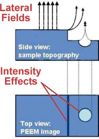
If the sample has topographic features – steps or trenches –
boundary conditions on the applied field result in locally strong
lateral fields at the sample surface.
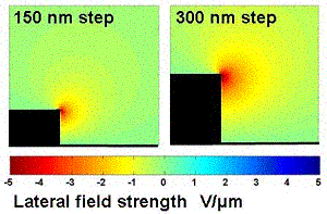
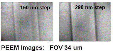
When emitted, the electrons are traveling quite slowly so that these short-range lateral fields can have an unexpectedly large effect on the electrons’ flight. The field kicks electrons away from the step edge. In the image, this results in a dark edge. Our calculations of the lateral field strength indicate that the range of these fields is comparable to step height, so the effect should be greater for a taller step. |
|
Test Sample Structure
|
 |
|
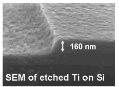
| This increasing edge effect is apparent in PEEM images of steps etched in Titanium. At left is an SEM image of a typical step profile. On the right, PEEM images of these same steps show the darker edge effect for the taller step. | ||
|
 |
|

